
I joke about me being too tall to see slime molds down there on the forest floor, but occasionally I get to spot them. Then comes the process of setting up my camera and tripod in the most awkward position possible and holding my breath for long exposures… I guess if macro photographers ever do yoga or free-diving they will be a force to reckon with in these disciplines.
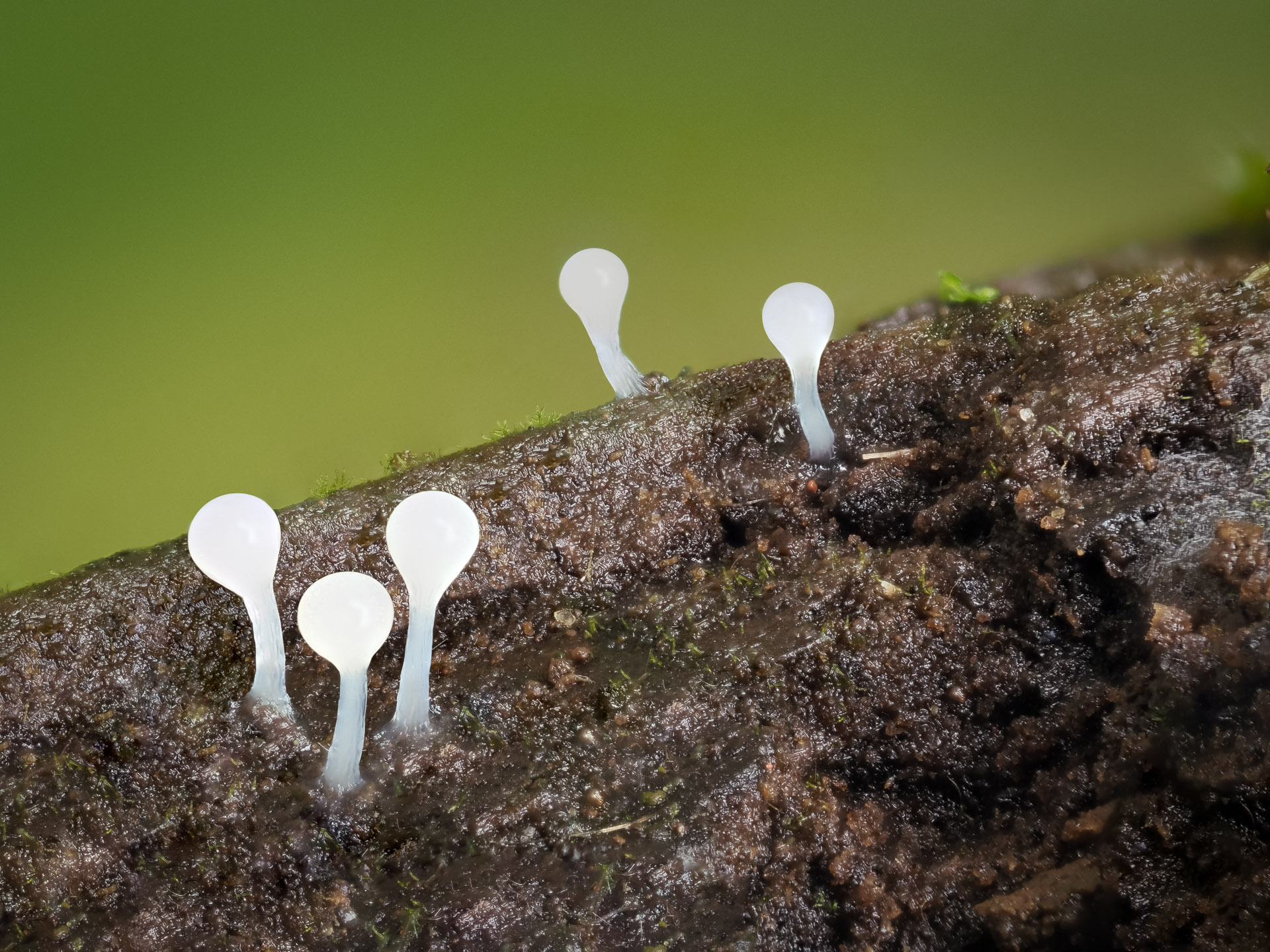
There are three phases in the life cycle of slime molds that are very photogenic: The unopened fruiting bodies or sporangia, the equivalent of mushrooms; the already opened sporangia that held the spores; and the viscous liquid phase that crawls about, looking for nutrients. I think the latter one looks better in stop motion videos though.
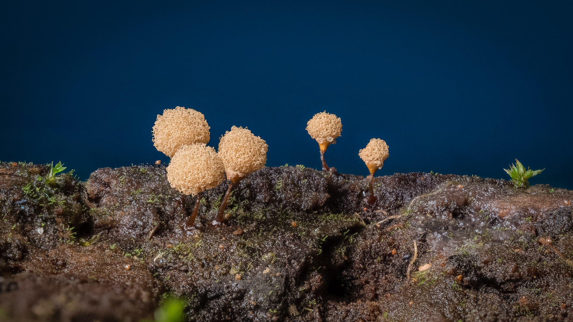
These things are about a millimeter tall and require stacking macro images to get a good looking image. Luckily, my OM System OM-1 camera can do this automatically as long as I don’t need to do stack more than 15 photos. If more images are needed the camera will take as many as I want but then I have to stack them later using Helicon Focus. Either way it saves you from having to slowly slide the camera in a focusing rail to get each image for the stack.
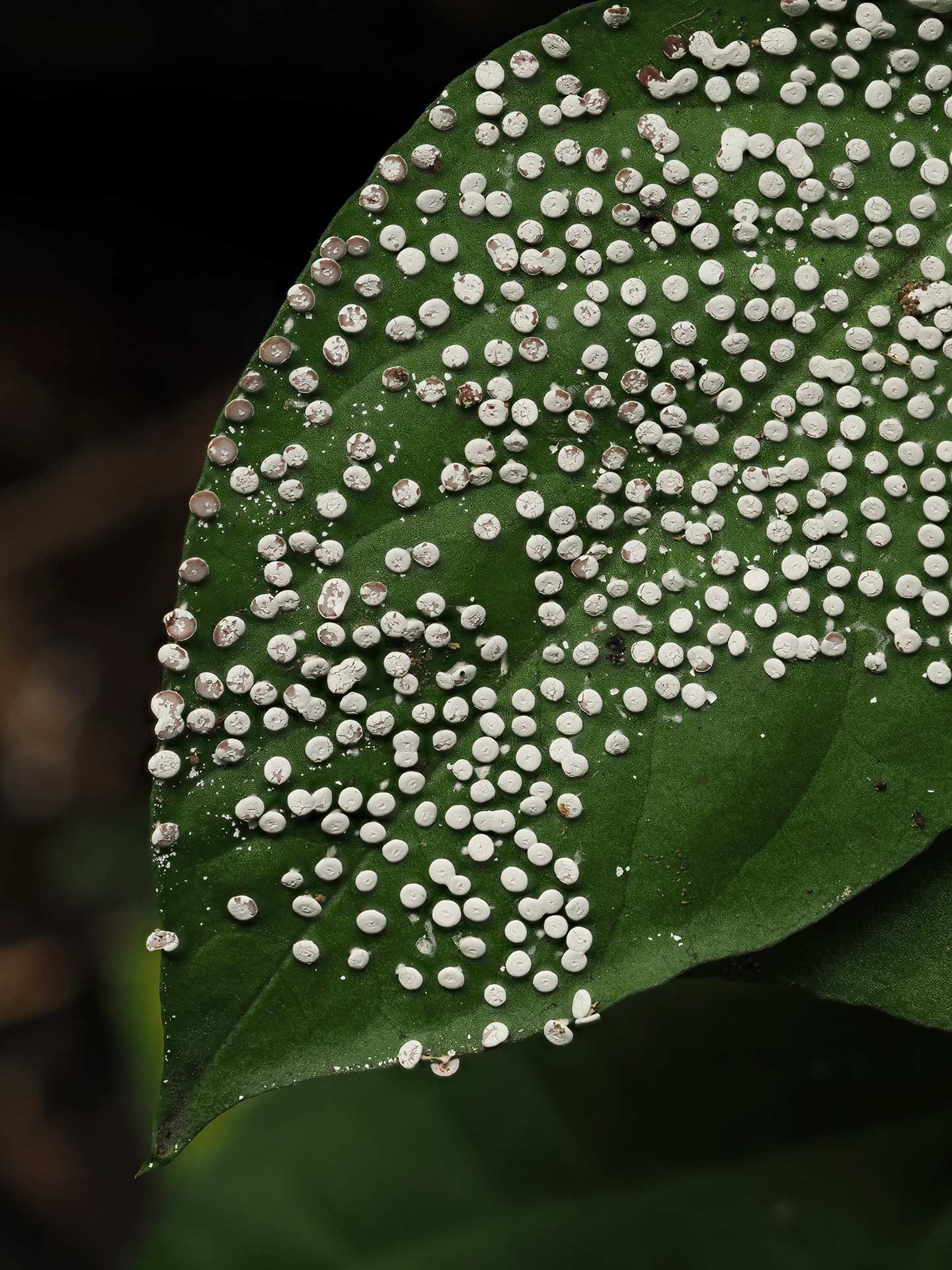
The white Didymium growing on a leaf was an in-camera stack. You get a ready to use stacked jpeg and also saves the individual raw files in case you need them. The other ones are stacked using Helicon.
Finding a nice subject among these tiny organisms brings about a sense of discovery that certainly makes up for any technical hardships!

Fascinating slime moulds! Have you see time lapse footage?
LikeLike